SOX10 and HaloPROTACs: Beyond Gene Silencing
Drug Discovery News · Oct 27, 2026 · Targeted Protein Degradation · 6 min read
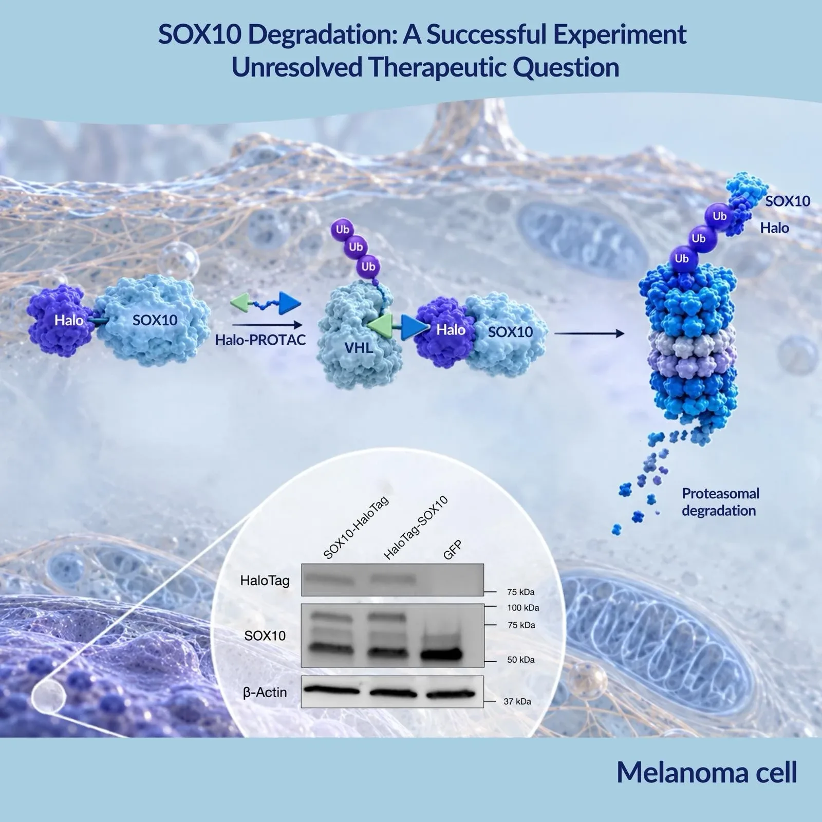
A bioRxiv preprint from Purvis, Slavoff and colleagues at Yale uses a HaloTag fusion and the commercial degrader HaloPROTAC3 to remove SOX10 from patient-derived melanoma cells and to map the genes that depend on it. It shows what a tag-degrader can establish before a degrader for the native protein exists, and where that evidence stops: the data describe an engineered, overexpressed SOX10–HaloTag fusion, not the druggability of native SOX10. The work is a preprint and has not been peer reviewed.
No pocket, and a knockout that stops growth
A degrader program normally starts from a ligand, and high-affinity ligands are lacking for most human proteins (Tovell et al., 2019). The authors note that SOX10 is predominantly intrinsically disordered outside its HMG DNA-binding domain. Genetic loss is also hard to interpret: silencing or deletion abolishes melanoma cell proliferation, so direct effects of SOX10 depletion are difficult to separate from those of the resulting dormancy (Purvis et al., 2026). SOX10 loss also gives rise to invasive, drug-tolerant cells (Capparelli et al., 2022). The authors therefore remove the protein on demand with a degrader.
Swapping endogenous SOX10 for a degradable copy
The study used WW165, an early-passage, patient-derived cutaneous melanoma line carrying BRAF V600K. HaloTag was fused to either end of SOX10, and both fusions were degraded by HaloPROTAC3, the VHL-recruiting compound of the original series (Buckley et al., 2015). The VHL-nonbinding stereoisomer ent-HaloPROTAC3 did not support degradation, and the proteasome inhibitor epoxomicin blocked it.
The authors then deleted the endogenous SOX10 alleles with inducible Cas9, using a codon-optimized transgene, and isolated a monoclonal line expressing only the C-terminal fusion. This line grew slightly more slowly than parental cells and expressed the fusion at about seven times the endogenous SOX10 level. At 1.25 µM, HaloPROTAC3 reduced the fusion by about 67% at 24 h and about 95% at 48 h; at 72 h it was undetectable by Western blot. Treatment inhibited proliferation of the engineered line but not of parental cells.
From 707 genes to two validated targets
After 48 h of HaloPROTAC3, RNA-seq (adjusted P < 0.01, log2 fold change ≤ −2) identified 707 downregulated genes, including the established SOX10 targets MITF, ERBB3 and DCT. Intersection with genes that lost chromatin accessibility in ATAC-seq left 101 candidates that depend on SOX10, directly or indirectly.
In parental cells, siRNA reduced SOX10 by about 73% and gave 571 downregulated genes at the same thresholds. Of these, 282 overlapped with the degradation set, and fold changes correlated (coefficient 0.66). Most of the 425 genes called only after degradation were also lower after siRNA but below the cutoff, which the authors speculate reflects more complete or rapid depletion. Three features of the design are useful context. The two perturbations used different cells and different starting SOX10 levels, so residual protein is not directly comparable. Expression was profiled once, at 48 h, so the list includes primary and secondary effects. And the RNA-seq compares HaloPROTAC3 with vehicle; adding the ent-HaloPROTAC3 control would help separate loss of SOX10 from covalent occupancy of its tag.
From the 101 genes, the authors chose 13 not previously reported as SOX10 targets. They report RT-qPCR confirmation for nine and loss of protein for five, including NAV2, NCKAP5 and CD200; for six genes no specific antibody or band was obtained. SOX10 ChIP-seq from a different melanoma line, 501Mel (Laurette et al., 2015), shows promoter-proximal peaks at ten of the 13. The authors then looked beyond WW165: in published RNA-seq from hundreds of sun-exposed and sun-shielded melanomas, stratified into 275 lines with normal SOX10 expression and 22 with low, nine of the 13 genes tracked SOX10 levels. Integrating every level, they report NAV2 and NCKAP5 as fully validated SOX10 targets, and TDRD7 as supported by all levels except the Western blot, for which no specific antibody was available. The evidence is convergent but correlative; it does not show that SOX10 binding drives their transcription in WW165 cells.
Linkers, ligases and a covalent bond
Buckley et al., 2015 introduced HaloPROTAC3, with a DC50 of 19 ± 1 nM and 90% maximal degradation of a GFP–HaloTag7 reporter in HEK293 cells. Tovell et al., 2019 optimized the VHL ligand (HaloPROTAC-E: DC50 3–10 nM, about 95% depletion at 48 h) and tagged SGK3 and VPS34 at their endogenous loci by CRISPR knock-in; they noted that all earlier HaloPROTAC work had used overexpressed proteins, as the SOX10 study does. CRBN-based versions followed. Ody et al., 2023 reported higher activity than VHL-based HaloPROTACs against an EGFP–HaloTag fusion at sub-micromolar concentrations, and Stevens et al., 2026 found dihydrouracil CRBN binders more potent and selective than IMiD-based analogues among more than 100 compounds in a FAK-tagged line. Each comparison was made in one test system and is not a general ranking.
Linker length and the mode of target engagement matter for interpretation. In the original series, shorter linkers (HaloPROTAC5–7) gave less than 20% degradation, while longer ones were less effective than HaloPROTAC3 and showed auto-inhibition at higher concentrations; cooperativity was proposed as the explanation but not measured. Because the chloroalkane reacts covalently with HaloTag, complete degradation requires stoichiometric occupancy of the tagged protein, whereas PROTACs with non-covalent ligands can act catalytically (Tovell et al.). That is relevant for a fusion expressed at several times the endogenous level, and the preprint's main text does not discuss it.
Where the tag ends and SOX10 begins
The fusion is not native SOX10. It was expressed at about seven times the endogenous level and only partially rescued proliferation, as the authors note. Degrading the tagged protein also does not show that unmodified SOX10 can be degraded. The premise of a missing ligand needs qualification here: DrugMap identified a cysteine-reactive covalent SOX10 ligand that increases SOX10–SOX10 interactions and disrupts melanoma transcriptional signaling (Takahashi et al., 2024). The preprint's authors suggest converting such a ligand into a PROTAC and state that no PROTAC targeting endogenous SOX10 has been reported to date. It cites no data on whether the ligand can support a productive ternary complex.
Platform choice is model-dependent. Bondeson et al. compared five conditional degron tags across 16 targets; none worked efficiently for all, but at least one did for each. Magtoto et al. reported superior degradation by dTAG molecules over HaloPROTAC and NanoTAC in a reporter mouse. Neither supports a universal ranking.
SOX10 biology cuts both ways. Capparelli et al. found that SOX10 loss gives rise to invasive, drug-tolerant cells and that cIAP1/2 inhibitors selectively kill SOX10-deficient cells. A proliferation arrest in culture therefore cannot settle whether SOX10 degradation would benefit patients.
From a phenotype to a ligand
A tag-degrader experiment answers a bounded question: what happens in this cell line, with this fusion, when the protein is removed quickly and almost completely? A clear phenotype is a reason to invest in ligand discovery. It is not evidence that a ligand, once found, will support degradation, or that the phenotype will persist at native expression levels or in tumors.
For SOX10, the authors' proposed next steps are an optimized fusion and linker, another degron system, or conversion of the covalent ligand into a degrader. All three run through the same chemistry: structurally diverse E3 ligands, modular linkers and matched compound series in which one element changes at a time, as in the direct-to-biology campaign of Stevens et al., 2026 — the bifunctional building blocks and linkers that research organizations such as Enamine Germany supply. Which of the three ends in a molecule that degrades native SOX10 is still an open question.
Sources
Enamine Germany