A 100,000-Fold Difference: How Linker Geometry Transforms Bivalent EGFR Inhibitors
Drug Discovery News · Oct 31, 2026 · Fragment-Based Drug Discovery · 5 min read
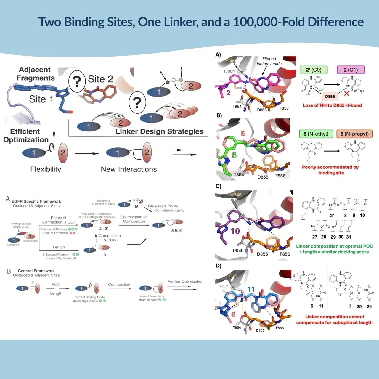
Connecting two molecules that bind neighboring pockets of the same protein seems like a straightforward way to improve potency. Each fragment already has its own binding interactions, so combining them should produce a stronger inhibitor. In practice, the connection can interfere with both.
A recent study by Patel and colleagues in the Journal of Medicinal Chemistry shows just how dramatic this effect can be. By changing the attachment point, length, and chemical composition of linkers in bivalent EGFR inhibitors, the researchers observed differences in biochemical potency exceeding five orders of magnitude. Their findings help explain why fragment linking can produce exceptionally potent compounds, or fail despite promising starting fragments.
Two Binding Sites, One Molecular Challenge
EGFR is an established therapeutic target in non-small cell lung cancer. Mutations such as L858R and T790M have driven the development of inhibitors that recognize different regions of its kinase domain.
Among these approaches are ATP-allosteric bivalent inhibitors (AABIs). They combine an ATP-competitive fragment with an allosteric fragment that occupies a neighboring pocket. Both binding elements are incorporated into one molecule, allowing simultaneous engagement of two distinct regions of EGFR.
Patel and colleagues studied inhibitors containing an imidazole-based ATP-site binder and a dibenzodiazepinone-derived allosteric binder. The challenge was to connect these fragments without disturbing the interactions that made them effective individually.
The researchers systematically varied three elements of linker architecture: the point of connection (POC), the spacer length, and its chemical composition. Biochemical assays against EGFR L858R/T790M were combined with X-ray crystallography, molecular docking, and molecular dynamics simulations to explain the resulting structure–activity relationships.
The largest differences came from where and how the two fragments were connected.
Moving the Attachment Point Changes Everything
The position of linker attachment had an extraordinary effect on EGFR inhibition.
Connecting the linker through the C9 position of the allosteric fragment produced highly potent inhibitors. Moving the attachment to C1 reduced biochemical potency by approximately 500-fold. Other attachment positions were even less favorable.
The contrast between previously reported N-linked inhibitors and optimized C-linked analogues was greater still, exceeding 100,000-fold in biochemical potency.
The structural explanation is particularly interesting. Although the ATP-binding fragment can retain its interactions with the kinase, changing the attachment point influences how the allosteric fragment fits into its pocket.
For the C1-linked analogue, molecular docking suggests that the lactam group adopts a flipped orientation, losing an important hydrogen bond between its NH group and EGFR residue D855. The more favorable C9 connection allows the allosteric fragment to retain the interactions associated with its preferred binding mode.
The linker does not need to make a strong interaction with the protein itself. Its position determines whether the two connected fragments can engage their respective pockets without compromising each other's binding geometry.
When One Extra Carbon Makes the Difference
Changing linker length produced another striking example of the sensitivity of these molecules.
The original N-linked inhibitors contained a short methylene connection and showed weak biochemical activity. Extending the linker by one carbon produced compound 5, with a substantial improvement in potency.
X-ray crystallography explained the result. The ethylene linker allowed the allosteric dibenzodiazepinone fragment to recover a favorable conformation resembling that of the unlinked allosteric inhibitor. Adding another carbon to generate the propylene-linked compound 6 reduced potency again.
The relationship between linker length and activity was therefore far from linear. A longer connection did not necessarily provide a better fit or greater freedom to establish productive interactions.
Compound 5 also showed unusual biphasic inhibition behavior, with two apparent inhibitory components. The authors suggest that conformational differences may contribute, although the underlying mechanism remains unresolved. This complexity matters when interpreting the SAR. A substantial improvement in biochemical activity does not always mean that the molecule has adopted a single, well-defined binding behavior.
A 25 pM Inhibitor — and a Surprising Role for Linker Chemistry
Once the researchers identified favorable attachment geometry, they examined whether the chemical composition of the linker could provide further improvements.
A simple reversal of an amide group produced compound 8, the most potent newly synthesized inhibitor in the study, with a biochemical IC50 of approximately 25 pM against EGFR L858R/T790M. Its crystal structure revealed an intramolecular hydrogen bond between the linker carbonyl and the allosteric fragment. This interaction may help support the productive binding conformation.
However, the following experiments produced an unexpected result. Replacing the amide linkage with an amine or ether generated compounds 9 and 10, which retained similarly strong biochemical potency. The amide interaction was therefore not essential for maintaining exceptionally tight inhibition.
Once the attachment point and linker geometry were optimized, several chemically distinct connections could support productive binding. This tolerance creates opportunities for further molecular optimization. Amides, amines, and ethers differ in hydrogen-bonding properties, polarity, and chemical behavior. Exploring these alternatives can help adjust molecular properties without necessarily sacrificing biochemical potency.
The study also examined whether additional linker-mediated hydrogen bonds could rescue compounds with unfavorable spacer lengths. Although some modifications improved activity, they could not fully compensate for poor binding geometry. The order in which linker variables are optimized clearly matters.
Flexibility Without Losing Molecular Recognition
The crystallographic structures and molecular dynamics simulations reveal why the strongest inhibitors work. The C-linked compounds allow greater conformational freedom around the connection to the allosteric fragment. This enables the fragment to adapt to its pocket while preserving productive ATP-site engagement. The less favorable architectures restrict these movements or force the fragments into less suitable orientations.
Importantly, flexibility here does not simply mean increasing the number of rotatable bonds. The relevant property is the ability of the connected fragments to retain their preferred binding modes without the linker pulling either of them out of position. That is what the authors identify as the key determinant of potency, and it gives fragment linking a working order: settle the point of connection and the length first, and treat linker composition as the final adjustment rather than the starting point.
Sources
Enamine Germany