Delivering Cyclin K Degradation to HER2-Positive Cancer Cells
Drug Discovery News · Nov 20, 2026 · Targeted Protein Degradation · 8 min read
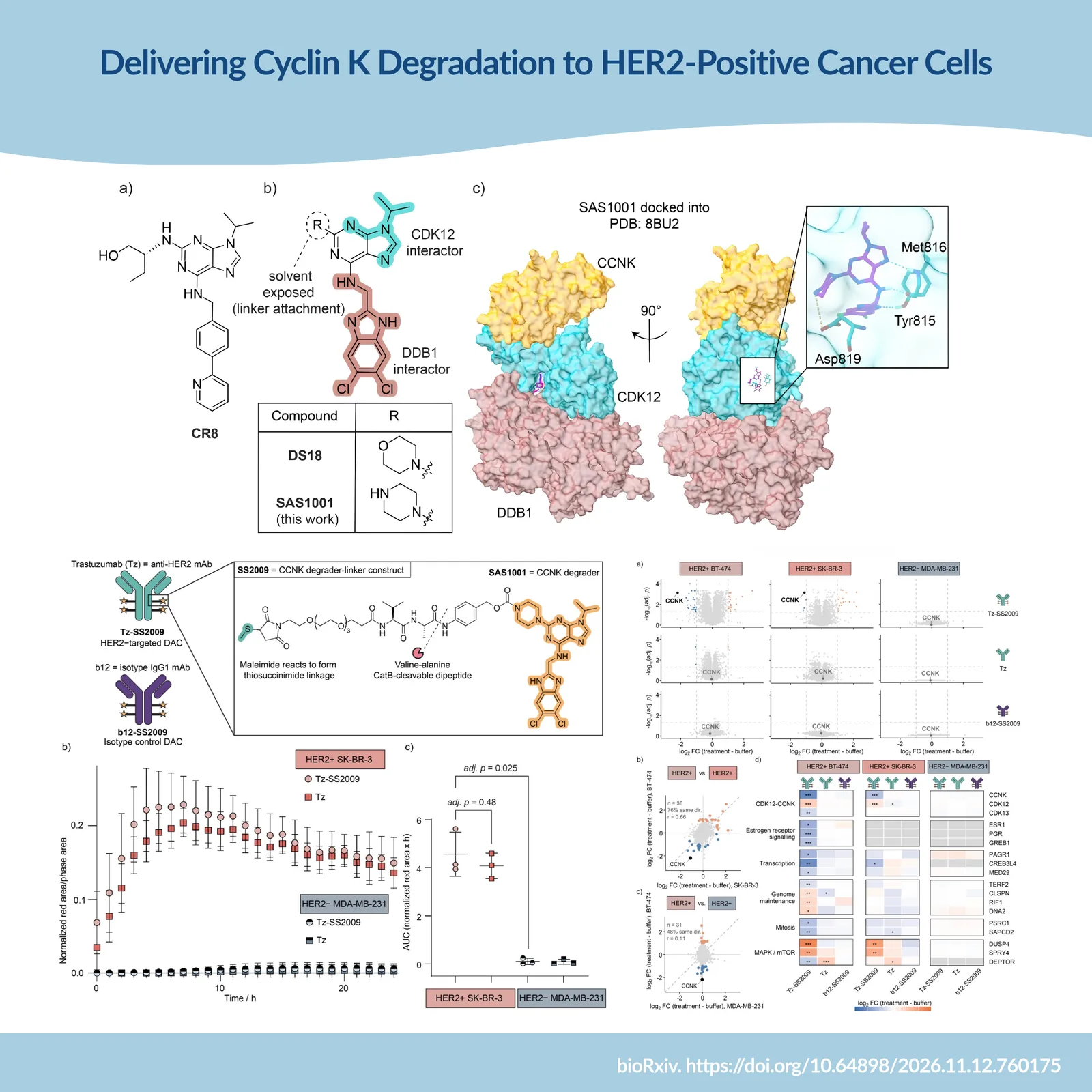
A cyclin K degrader killed HER2-negative cells at 10 nM. Four times as much of the same payload, delivered on an antibody, left them untouched. That contrast is the point of a new preprint from Imperial College London, EPFL and ADC Therapeutics. Cyclin K is potently degradable and broadly essential. Potency was never the problem — location was. For a molecular glue, the linker is the design problem. There is no spare binding element, so the attachment point has to be built into the scaffold from structural data — or the ternary complex stops forming. One morpholine-to-piperazine swap later, it survived conjugation to trastuzumab. Selectivity, it turns out, need not come from the small molecule at all.
Potency is rarely the limiting factor in targeted protein degradation any more. Sub-nanomolar degraders of previously intractable targets have become routine. The harder question is where degradation happens — because when the protein being removed is essential in healthy tissue as well as in tumor, a more potent degrader does not widen the therapeutic window. It becomes a more efficient way to deplete the target everywhere.
Cyclin K (CCNK) is a textbook case. As the obligate partner of CDK12 and CDK13 — kinases sharing over 93% identity across the kinase domain, which has frustrated selective inhibitor work for years — removing the cyclin disables both active complexes at once. That is what makes degradation attractive, and what makes it risky: CCNK is broadly expressed, functionally essential, and has proposed kinase-independent roles that would broaden the consequences of systemic depletion.
A new study by Srdanovic and colleagues at Imperial College London, EPFL and ADC Therapeutics asks whether an antibody can supply what the small molecule cannot. The group conjugated a potent CCNK molecular glue degrader to trastuzumab, testing whether cellular selectivity can come from the delivery system instead.
Why HER2 is an unusually good fit. The pairing is not arbitrary. CDK12 sits on the 17q12 amplicon and is frequently co-amplified with ERBB2, the gene encoding HER2 — a co-amplification associated with more aggressive phenotypes and metastatic progression. Roughly 15–20% of breast cancers are HER2-positive, HER2 is a clinically validated internalizing antigen with a mature conjugate toolkit, and CDK12 has been implicated in trastuzumab resistance, with CDK12 inhibition restoring trastuzumab sensitivity in preclinical models. The antigen and the target dependency travel together in the same tumors - an unusually clean rationale for a delivery strategy.
For a molecular glue, linker placement is the design problem. A PROTAC offers two separable binding elements and, with them, reasonably forgiving exit vectors. A molecular glue offers neither. Its activity depends on stabilizing an induced interface between two proteins, and a substituent that looks peripheral by conventional SAR can still alter the geometry or energetics of that interface. A viable attachment point must preserve not binding but productive ternary complex assembly.
That constraint explains why degrader–antibody conjugates have been dominated by cereblon-based glues: IMiD chemotypes come with well-mapped SAR and attachment points already exploited in CRBN-recruiting PROTACs. Convenience carries a liability, though. CRBN loss is tolerated in cultured cells, in mice and even in humans, making it a plausible resistance route — whereas DDB1, the adaptor exploited by CCNK glues, is broadly essential. Before this study, a non-CRBN glue had been delivered on an antibody-like scaffold only once, using CR8 on a HER2 affibody that binds its antigen covalently and releases payload by disulfide exchange.
A degrader designed to be conjugatable. The scaffold traces back to Słabicki and colleagues' discovery that CR8, characterized as a CDK inhibitor, degrades CCNK through an unexpected glue mechanism: it promotes an interaction between the CDK12–CCNK complex and DDB1, adaptor of the CUL4 ligase, positioning CCNK for ubiquitylation. Kozicka and colleagues then mapped which modifications preserve productive complex formation — including that the 2-position is largely solvent-exposed.
Srdanovic and colleagues used that insight deliberately. Taking the optimized CR8 derivative DS18, they replaced its morpholine with a piperazine, installing a conjugation-ready amine where one should be tolerated. Docking into the DDB1–CDK12–CCNK complex indicated a retained binding pose, plus a predicted salt bridge between the protonated piperazine and Asp819.
The resulting compound, SAS1001, did not merely survive the modification:
• Ternary complex formation (TR-FRET): EC₅₀ 3.3 nM, versus 8.4 nM for CR8 assayed in parallel.
• Reporter degradation: DC₅₀ 0.92 nM, versus 13 nM for CR8 and 6.0 nM for SR-4835.
• Endogenous CCNK across four breast cancer lines: apparent DC₅₀ 32–97 pM, with Dmax above 99% in SK-BR-3, BT-474 and MDA-MB-231, and 83% in MCF-7.
• Mechanism: degradation was abolished by neddylation, E1 and proteasome inhibition.
• Selectivity: at 6 h, CCNK was one of only two altered proteins among 7,270 quantified in SK-BR-3, and one of eleven among 7,599 in MDA-MB-231.
Reporter and endogenous values describe different systems and are not interchangeable. The design conclusion holds regardless: a structure-guided modification produced a conjugation handle without sacrificing the degradation mechanism — a deliberate result, not an opportunistic one. Crucially, the free degrader also demonstrated the problem it was meant to address. SAS1001 inhibited proliferation with IC₅₀ values of 1.0–14 nM across the panel — but only 1.7-fold (SK-BR-3) and 3.8-fold (BT-474) more potently than in HER2-negative MDA-MB-231 cells, and degradation proceeded at least as fast in the HER2-negative line at every timepoint measured. Potent degradation, essentially no cellular selectivity.
From payload to conjugate. The linker–payload construct SS2009 combines a PEG spacer, a cathepsin B-cleavable valine–alanine dipeptide and a self-immolative PABC group designed to release unmodified SAS1001. Incubation with human liver cathepsin B confirmed cleavage and traceless release. SS2009 was conjugated to trastuzumab (Tz-SS2009) and, as a non-targeting control, to b12, an anti-gp120 IgG1 recognizing no human cell-surface antigen (b12-SS2009). Both conjugates reached an average drug-to-antibody ratio of approximately 4.
That matched control is what makes the rest of the study interpretable. Antibody conjugates can enter cells by antigen-independent routes, and catalytic payloads can produce biology even at inefficient uptake — so an advantage over a payload- and DAR-matched non-targeting conjugate is far more informative than comparison with untreated cells. The same laboratory established this discipline earlier with a trastuzumab–BRD4 PROTAC conjugate; the glue payload is the new variable.
Real-time imaging confirmed preferential uptake of Tz-SS2009 in HER2-positive SK-BR-3 over HER2-negative MDA-MB-231 cells — and uptake indistinguishable from unconjugated trastuzumab, indicating that the hydrophobic payload does not impair receptor-mediated internalization.
What the antibody actually buys. In HER2-positive cells, Tz-SS2009 degraded CCNK with DC₅₀ values of approximately 270 pM (BT-474) and 210 pM (SK-BR-3) in antibody equivalents — roughly 1 nM in payload equivalents at DAR 4.
The selectivity figures are substantial:
• 45-fold lower CCNK abundance in BT-474 cells with Tz-SS2009 than with isotype-matched b12-SS2009 at 10 nM (8.6-fold at 1 nM).
• 51-fold lower CCNK abundance in HER2-positive BT-474 than in HER2-negative MDA-MB-231 under Tz-SS2009.
• 13-fold (SK-BR-3) and 36-fold (BT-474) antiproliferative selectivity relative to MDA-MB-231 — an 8- to 10-fold improvement over the unconjugated degrader.
Two further results deserve emphasis, because they address the obvious objections. First, the trastuzumab confound. In SK-BR-3 cells Tz-SS2009 gave an IC₅₀ of 14 nM against 200 nM for the non-targeting conjugate, while unconjugated trastuzumab never reached 50% inhibition up to 500 nM — so that window belongs to the delivered degrader. In BT-474 cells trastuzumab is intrinsically active (IC₅₀ 5.9 nM versus 5.0 nM for the conjugate), and the authors say plainly that attribution is confounded. Reporting both rather than only the favorable case is the right call. Second, and most persuasive: 10 nM free SAS1001 killed MDA-MB-231 cells within 24 h, so comprehensively that proteomics was impossible. At DAR 4, 10 nM Tz-SS2009 carries 40 nM of the same payload — four times as much — yet those cells stayed healthy and yielded the deepest proteome of any condition analyzed (7,824 protein groups). The payload was present and did nothing, because it was never delivered.
Proteomics reinforced this. Responses to Tz-SS2009 in the two HER2-positive lines agreed directionally for 76% of changed proteins, while agreement with MDA-MB-231 fell to 48% — chance level, meaning the response in HER2-negative cells was absent rather than weaker. The isotype control altered no protein in any line. Downstream changes tracked known CDK12–CCNK biology: transcriptional regulation, genome maintenance, mitotic progression and estrogen signaling.
What the study does not establish. The honest limitations are as instructive as the results. Per payload equivalent, the conjugate is roughly 25- to 30-fold less potent than free SAS1001. Internalization, endolysosomal trafficking, linker cleavage, payload release and nuclear access each take a toll — and the basic piperazine installed for conjugation may itself invite lysosomal sequestration. The handle that made the molecule conjugatable may also limit it.
Selectivity is relative, not absolute. At the highest concentrations both conjugates reduced viability in HER2-negative cells, consistent with low-level antigen-independent uptake or premature linker cleavage — an effect a catalytic mechanism amplifies. CDK12 abundance also rose under conjugate treatment while falling with the free compound: plausibly exposure-dependent, but unresolved.
Most importantly, cellular selectivity is not a therapeutic index. CCNK depletion in healthy tissue remains the central risk, and only pharmacokinetic, biodistribution, efficacy and tolerability studies in vivo can address it. Clinical precedent is thin: the two GSPT1-directed conjugates to reach the clinic have both been discontinued. Nor is response likely to track HER2 expression alone — antigen accessibility, internalization kinetics, payload release and a tumor's actual dependence on CDK12–CCNK signaling will all contribute.
The chemistry sits on the critical path. The broader lesson is that molecular glue discovery and molecular glue delivery cannot be treated as separate disciplines. A scaffold can be exceptionally potent and entirely unsuitable for conjugation, if its accessible positions are the ones holding the ternary complex together. Conversely, one structure-guided substitution can create a viable attachment point while preserving degradation. The decision is made at the scaffold rather than at the linker, and it is made from structural data rather than discovered afterwards.
That reframes the practical work: structural characterization of glue complexes becomes a design input for conjugation-ready analogues, and progress depends on systematically varying attachment points, physicochemical properties and release chemistry. Each variant is a multi-step synthesis carrying a late-stage traceless-release constraint, made cleanly enough to interpret the biology that follows. Chemistry execution is not a downstream service here — it is where the strategy succeeds or fails.
Srdanovic and colleagues have not produced a treatment for HER2-positive cancer, and they do not claim to. What they have demonstrated is narrower and more useful: the cellular selectivity of a molecular glue need not come from the small molecule at all. It can be engineered into the chemistry and biology of its delivery.
Sources
Enamine Germany